Loading…
Cardiologist-arrhythmologist · Ezgu Niyat clinic
Every heartbeat matters.
Sherzod Nasirovich Sabirov is a cardiologist-arrhythmologist and Head of the Arrhythmology Department at the Ezgu Niyat clinic. In cardiac surgery and arrhythmology since 2010, including 10 years at the Bakulev Center in Moscow.
About the doctor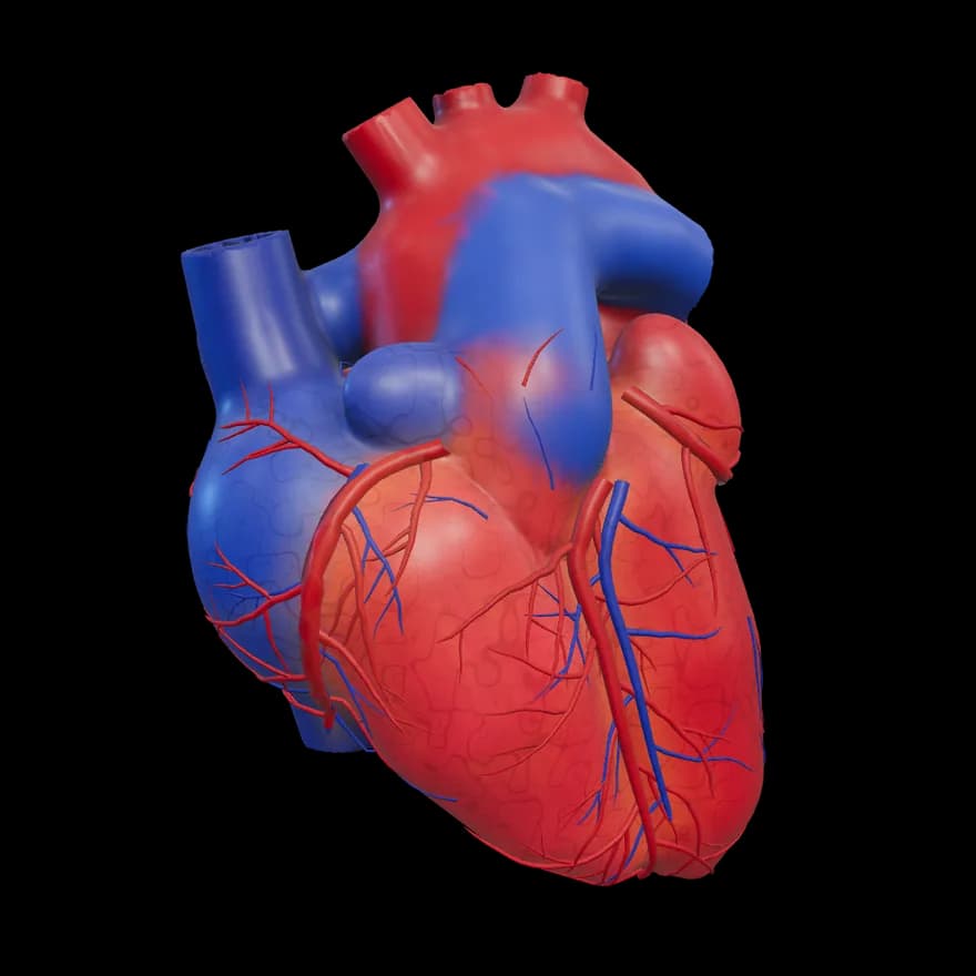
- 16 years
- in cardiac surgery and arrhythmologySince 2010: Bakulev Center; since 2020: Ezgu Niyat clinic
- 10
- years at the Bakulev CenterMoscow, 2010–2020: residency, PhD, cardiac surgery
- L. A. Bockeria
- student of the academicianSchool of Academician L. A. Bockeria
The information has been approved by the doctor.
What is bothering you?
Choose the topic that matches what you feel and read the general information. This is not a diagnosis — a decision requires a doctor's examination.
- The heart races
- The heart beats irregularly and seems to “stop”
- The heart suddenly starts racing and suddenly stops
- Fainting or nearly fainting
- The heart seems to beat very slowly, weakness
- A smartwatch showed an irregular rhythm
- I have been diagnosed with atrial fibrillation
Emergency: if you have severe chest pain, severe shortness of breath or fainting, do not wait.
Call 103
Sherzod Nasirovich Sabirov
Extensive clinical experience. Clear communication.
After the Tashkent Medical Academy, he spent 10 years (2010–2020) at the A. N. Bakulev Center in Moscow, in the school of Academician L. A. Bockeria, going through residency, a PhD and cardiovascular surgery. Since 2020, he has headed the Interventional Arrhythmology Department at the Ezgu Niyat clinic. Member of ESC and EHRA.
Full profileServices
All- 01Arrhythmologist consultationAdvice from a doctor who specializes in heart rhythm disorders.
- 02Cardiologist consultationA doctor’s examination and advice on the cardiovascular system.
- 03Assessment of heart rhythm disordersAssessing the rhythm based on your symptoms and test results.
- 04ECGA test that records the heart’s electrical activity.
- 05Holter monitoringContinuous recording of the heart rhythm over 24 hours.
One heartbeat
How the electrical impulse travels through the heart
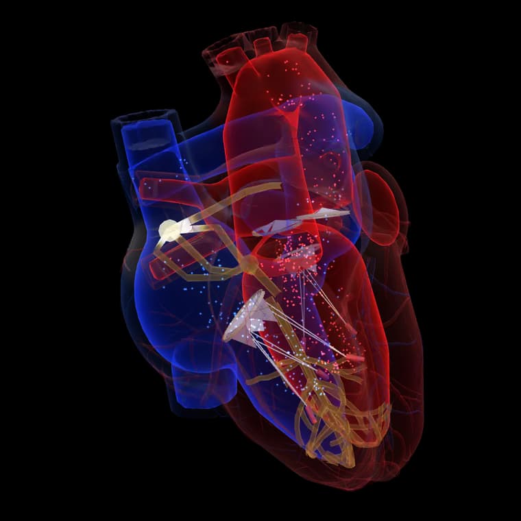
The impulse originates in the sinus node
The heart rhythm is set by the sinus node in the right atrium: it generates electrical impulses regularly.
The atria contract
The impulse spreads to the atria, and they push blood into the ventricles. On the ECG, this appears as the P wave.
A short pause in the atrioventricular node
The impulse is slightly delayed in the AV node — this leaves time for the ventricles to fill. On the ECG, this corresponds to the PQ interval.
The ventricles contract
The impulse passes to the ventricles through the bundle of His and the Purkinje fibers. On the ECG, this is the QRS complex.
Recovery and a new cycle
The ventricles recover electrically (T wave), and then the cycle starts again. A rhythm disorder can occur at any stage of this path — that is why the cause is determined by testing.
Educational illustration: the diagram is simplified and is not a diagnostic tool.
See for yourself
Understand the heart rhythm
In one cycle, the heart works in sequence: the atria contract, then the ventricles contract, and then the heart relaxes and fills with blood.
What do the colors mean?
Blue — pathways of oxygen-poor blood, red — oxygen-rich blood. Golden light — the electrical impulse spreading through the heart.
Blood path: venae cavae, right atrium, tricuspid valve, right ventricle, pulmonary artery, lungs, pulmonary veins, left atrium, bicuspid valve, left ventricle, aorta and the whole body.
Normal rhythm
about 60–100 beats/minThe spacing between beats is almost the same. Before each beat, the atrial signal (P wave) is visible.
Educational illustration: the line is not a real patient's ECG and is not a diagnostic tool.
Frequently asked questions
How do I book an appointment?
Press the “Book an appointment” button and leave your name and phone number. The administrator will contact you and arrange a convenient time. Sending a request does not mean the appointment is confirmed.
Do I need to write my symptoms in the form?
No. We recommend not writing information about your symptoms or diagnosis in a public form. You will give the details at the appointment or through the secure channel the administrator points you to.
What should I bring to the appointment?
Previous ECG, Holter and blood test results and a list of the medicines you are taking will be useful. More details are in the “For patients” section.
Is a diagnosis made through the site?
No. The site's materials and ECG illustrations are for educational purposes. A diagnosis is made only after a doctor's examination and tests.
Do not ignore your heart rhythm.
Leave your name and number — the administrator will contact you to arrange a convenient time.